38 fluorescent labels and light microscopy
Förster resonance energy transfer - Wikipedia In fluorescence microscopy, fluorescence confocal laser scanning microscopy, as well as in molecular biology, FRET is a useful tool to quantify molecular dynamics in biophysics and biochemistry, such as protein-protein interactions, protein–DNA interactions, and protein conformational changes. For monitoring the complex formation between two ... Western blot - Wikipedia Another method of secondary antibody detection utilizes a near-infrared fluorophore-linked antibody. The light produced from the excitation of a fluorescent dye is static, making fluorescent detection a more precise and accurate measure of the difference in the signal produced by labeled antibodies bound to proteins on a western blot.
Caveat fluorophore: an insiders’ guide to small-molecule ... Dec 23, 2021 · From its inception, fluorescence microscopy has been driven by advances in dyes. Early protein labels excited by ultraviolet (UV) light proved difficult to use for cellular imaging, and so Coons ...
Fluorescent labels and light microscopy
Fluorophore selection | Thermo Fisher Scientific - US Photostability in buffer is a relative measure of the percentage of initial fluorescence intensity remaining following 30 seconds of continuous illumination using a 40x/1.4NA objective and a 100-watt Hg-arc lamp as the light source with samples mounted in phosphate buffer; high photostability in buffer is ideal for live-cell imaging and detection Introduction to Fluorescent Proteins | Nikon’s MicroscopyU The brightness and fluorescence emission spectrum of enhanced yellow fluorescent protein combine to make this probe an excellent candidate for multicolor imaging experiments in fluorescence microscopy. Enhanced yellow fluorescent protein is also useful for energy transfer experiments when paired with enhanced cyan fluorescent protein (ECFP) or ... ZEISS Axioscope 5 Smart Laboratory Microscope Focus. Snap. Done. Forget about the 15 steps and clicks to document samples with multiple fluorescent labels. With Smart Microscopy, this is a thing of the past. Axioscope 5 with Axiocam 202 mono and Colibri 3 LED illumination take this workload from you. You keep your hands at the microscope stand. Relaxed.
Fluorescent labels and light microscopy. Fluorescence - Wikipedia Fluorescence is the emission of light by a substance that has absorbed light or other electromagnetic radiation.It is a form of luminescence.In most cases, the emitted light has a longer wavelength, and therefore a lower photon energy, than the absorbed radiation. ZEISS Axioscope 5 Smart Laboratory Microscope Focus. Snap. Done. Forget about the 15 steps and clicks to document samples with multiple fluorescent labels. With Smart Microscopy, this is a thing of the past. Axioscope 5 with Axiocam 202 mono and Colibri 3 LED illumination take this workload from you. You keep your hands at the microscope stand. Relaxed. Introduction to Fluorescent Proteins | Nikon’s MicroscopyU The brightness and fluorescence emission spectrum of enhanced yellow fluorescent protein combine to make this probe an excellent candidate for multicolor imaging experiments in fluorescence microscopy. Enhanced yellow fluorescent protein is also useful for energy transfer experiments when paired with enhanced cyan fluorescent protein (ECFP) or ... Fluorophore selection | Thermo Fisher Scientific - US Photostability in buffer is a relative measure of the percentage of initial fluorescence intensity remaining following 30 seconds of continuous illumination using a 40x/1.4NA objective and a 100-watt Hg-arc lamp as the light source with samples mounted in phosphate buffer; high photostability in buffer is ideal for live-cell imaging and detection
Light-Sheet Fluorescence Microscopy - 2020 - Wiley Analytical Science

Bacterias,Viruses and Parasites: Chlamydia trachomatis - Cause of STDs and Blindness.

Using single nuclei for RNA-seq to capture the transcriptome of postmortem neurons | Nature ...

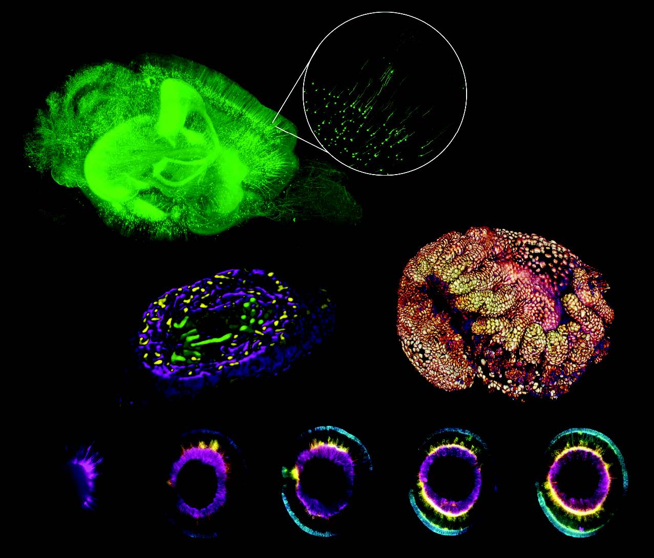
Light-sheet fluorescence microscopy helps reveal wiring in transparent brains | Laser Focus World

Fluorescence Microscopy - YouTube

Difference between Light Microscope and Electron Microscope (Light Microscope vs Electron ...
Microbiology Ch 1 Flashcards - Cram.com

Interactive Labeling of Fluorescence Microscopy Data - YouTube
Fluorescence Imaging

Scattering and fluorescent signal produced by unpurified NP-A-633 are... | Download Scientific ...

Part:BBa I714891:Experience - parts.igem.org

Confocal light absorption and scattering spectroscopic microscopy monitors organelles in live ...

Label-free prediction of three-dimensional fluorescence images from transmitted-light microscopy ...

Olympus FluoView Resource Center: Spectral Bleed-Through Artifacts in Confocal Microscopy

A schematic diagram of the light path in the ptychographic microscope... | Download Scientific ...
Post a Comment for "38 fluorescent labels and light microscopy"