43 light microscope with labels
Solved Label the image of a compound light microscope ... Experts are tested by Chegg as specialists in their subject area. We review their content and use your feedback to keep the quality high. Transcribed image text: Label the image of a compound light microscope using the terms provided. Cell Membrane Under Light Microscope Labeled : Functions ... The structures within the cell are referred to as organelles. NCERT RD Sharma Cengage KC Sinha. Some of the cell organelles that can be observed under the light microscope include the cell wall, cell membrane, cytoplasm, nucleus, vacuole and chloroplasts.
Labelled Diagram Of A Light Microscope | Products ... Products/Services for Labelled Diagram Of A Light Microscope Microscopes - (706 companies) ...and transmission electron microscopes. Acoustic and ultrasonic microscopes use sound waves to create images of the sample. Compound microscopes use a single light path. These types of microscopes can have a single eyepiece (monocular) or a dual eyepiece...
Light microscope with labels
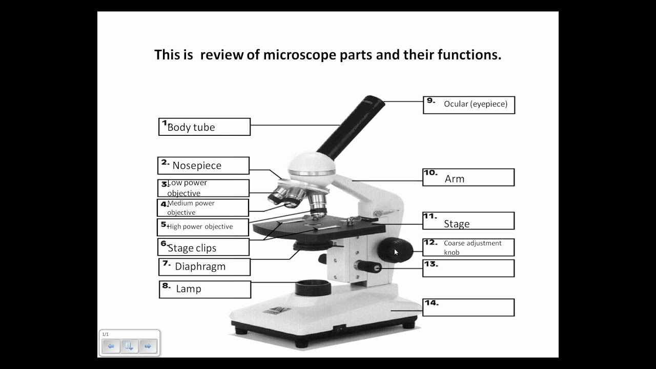
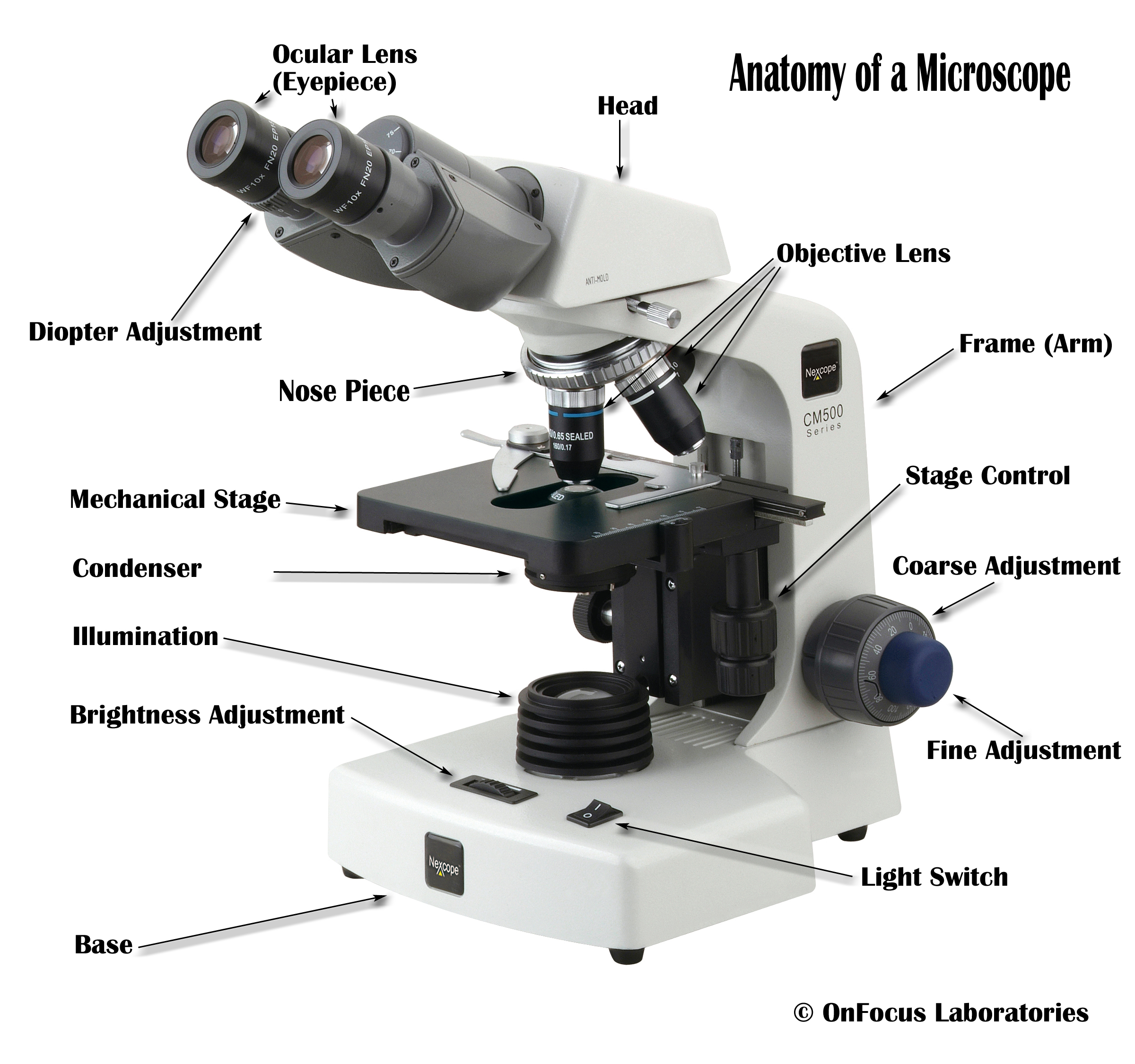
Parts of a microscope with functions and labeled diagram Apr 19, 2022 · Head – This is also known as the body. It carries the optical parts in the upper part of the microscope. Base – It acts as microscopes support. It also carries microscopic illuminators. Arms – This is the part connecting the base and to the head and the eyepiece tube to the base of the microscope. Microscope Parts and Functions With Labeled Diagram and ... Microscope Parts and Functions With Labeled Diagram and Functions How does a Compound Microscope Work?. Before exploring microscope parts and functions, you should probably understand that the compound light microscope is more complicated than just a microscope with more than one lens.. First, the purpose of a microscope is to magnify a small object or to magnify the fine details of a larger ... Label the microscope — Science Learning Hub All microscopes share features in common. In this interactive, you can label the different parts of a microscope. Use this with the Microscope parts activity to help students identify and label the main parts of a microscope and then describe their functions. Drag and drop the text labels onto the microscope diagram.
Light microscope with labels. A quick guide to light microscopy in cell biology - PMC Laser-scanning confocal microscopy is excellent for rejecting out-of-focus light and acquiring 3D image data, but in general it is less sensitive and more phototoxic than spinning-disk confocal microscopy. For live specimens for which 3D information is required, spinning-disk confocal microscopy should be considered. Labeling the Parts of the Microscope | Microscope activity ... Description A collection of microscope diagrams and worksheets for science class. Download them all in one convenient PDF, and select the version that's best for your classroom. This PDF contains the following: 1. Parts of a Microscope Diagram - Color 2. Parts of a Microscope Diagram - Black and White 3. Simple Microscope - Diagram (Parts labelled), Principle ... A simple microscope consists of Optical parts Mechanical parts Labeled Diagram of simple microscope parts Optical parts The optical parts of a simple microscope include Lens Mirror Eyepiece Lens A simple microscope uses biconvex lens to magnify the image of a specimen under focus. Microscope, Microscope Parts, Labeled Diagram, and Functions Revolving Nosepiece or Turret: Turret is the part of the microscope that holds two or multiple objective lenses and helps to rotate objective lenses and also helps to easily change power. Objective Lenses: Three are 3 or 4 objective lenses on a microscope. The objective lenses almost always consist of 4x, 10x, 40x and 100x powers. The most common eyepiece lens is 10x and when it coupled with ...
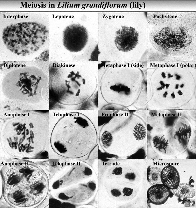
Microscope Labeling - The Biology Corner Students label the parts of the microscope in this photo of a basic laboratory light microscope. Can be used for practice or as a quiz. Name_____ Microscope Labeling . Microscope Use: 15. When focusing a specimen, you should always start with the _____ objective. Animal Cell Under Light Microscope Labelled : Draw and ... Most cells are visible under a light microscope, but mitochondria and bacteria are barely visible. Record the microscope images using labelled diagrams or produce digital images. We say cells are microscopic because they can only be seen under a microscope. Note the whiplike flagellum that gives the cell a threadlike appearance. Compound Microscope Parts, Functions, and Labeled Diagram ... Compound Microscope Definitions for Labels. Eyepiece (ocular lens) with or without Pointer: The part that is looked through at the top of the compound microscope. Eyepieces typically have a magnification between 5x & 30x. Monocular or Binocular Head: Structural support that holds & connects the eyepieces to the objective lenses. Parts of the Microscope with Labeling (also Free Printouts) Mar 07, 2022 · Parts of the Microscope with Labeling (also Free Printouts) A microscope is one of the invaluable tools in the laboratory setting. It is used to observe things that cannot be seen by the naked eye. Table of Contents 1. Eyepiece 2. Body tube/Head 3. Turret/Nose piece 4. Objective lenses 5. Knobs (fine and coarse) 6. Stage and stage clips 7. Aperture
Microscope labels Flashcards | Quizlet Microscope label Learn with flashcards, games, and more — for free. Microscope Drawing And Label at PaintingValley.com ... Tags: microscope, label All rights to paintings and other images found on PaintingValley.com are owned by their respective owners (authors, artists), and the Administration of the website doesn't bear responsibility for their use. Light Microscope: Functions, Parts and How to Use It ... Compound light microscope: has a higher magnification than a simple microscope because it uses at least two sets of lenses, an objective lens and an eyepiece. Light Microscope Function. The function of the light microscope is based on its ability to focus a beam of light through a very small and transparent specimen, to produce an image. Microscope Labeling - The Biology Corner 1) Start with scanning (the shortest objective) and only use the COARSE knob . Once it is focused… 2) Switch to low power (medium) and only use the COARSE knob . You may need to recenter your slide. Once it is focused.. 3) Switch to high power (long objective).
Search in gallery
Labeling the Parts of the Microscope | Microscope World ... Labeling the Parts of the Microscope This activity has been designed for use in homes and schools. Each microscope layout (both blank and the version with answers) are available as PDF downloads. You can view a more in-depth review of each part of the microscope here. Download the Label the Parts of the Microscope PDF printable version here.
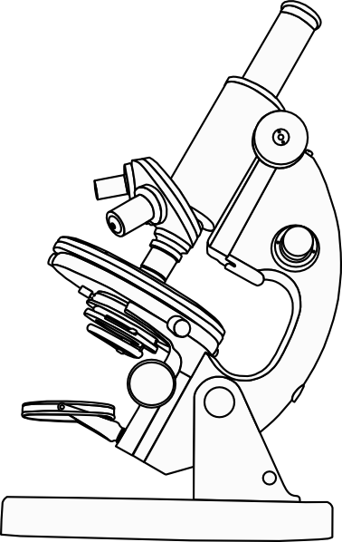
Microscope Clip Art at Clker.com - vector clip art online, royalty free & public domain
Microscope Labeling Game - PurposeGames.com This is an online quiz called Microscope Labeling Game. There is a printable worksheet available for download here so you can take the quiz with pen and paper. This quiz has tags. Click on the tags below to find other quizzes on the same subject. Science.
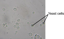
Yeast Bubbles - Experiments on Microscopes 4 Schools
Required practical - using a light microscope - Cells in ... Care must be taken when handling coverslips and microscope slides. Drawing the image Record the microscope images using labelled diagrams or produce digital images. When first examining cells or...
Microscope Review.wmv - YouTube
An Introduction to the Light Microscope, Light Microscopy ... For example, using green light with a wavelength of 550 nm and an objective with a typical NA of 0.7, a standard light microscope can resolve features down to a limit of 0.61 × (550 nm)/0.7 ≈ 480 nm, which is sufficient to observe cells (typically 10 µm size), but not enough to observe details of smaller organisms, e.g. viruses (typically ...

Microscope World Blog: Carpet under the Microscope
PDF Parts of the Light Microscope - Science Spot F. LIGHT SOURCE Projects light UPWARDS through the diaphragm, the SPECIMEN, and the LENSES H. DIAPHRAGM Regulates the amount of LIGHT on the specimen E. STAGE Supports the SLIDE being viewed K. ARM Used to SUPPORT the B. NOSEPIECE microscope when carried Holds the HIGH- and LOW- power objective LENSES; can be rotated to change MAGNIFICATION.
Microscope Diagram to Print
Light Microscope- Definition, Principle, Types, Parts ... Nov 07, 2021 · A light microscope is a biology laboratory instrument or tool, that uses visible light to detect and magnify very small objects and enlarge them. They use lenses to focus light on the specimen, magnifying it thus producing an image. The specimen is normally placed close to the microscopic lens.

Microscope Clipart - Clipartion.com
Compound Microscope: Definition, Diagram, Parts, Uses ... Compound microscope is a type of optical microscope that is used for obtaining a high-resolution image. There are more than two lenses in a compound microscope. Learn about the working principle, parts and uses of a compound microscope along with a labeled diagram here.
Search in gallery
Microscope Types (with labeled diagrams) and Functions This is an advanced microscope that has specific application in viewing, observing and measuring the optical thickness and phase of completely transparent specimens and objects. A tiny interferometer is used and a specimen is placed on beam path of it. This path is split and then rejoined to create two superimposed images of the specimen in focus.
Post a Comment for "43 light microscope with labels"