39 microscope with labels and functions
5 Best Microscopes for Kids Reviews of 2021 - BestAdvisor.com The AmScope M30-ABS-KT2-W microscope is a real find for your little one to dive deeper into the world of science, discover new things, and practice self-development. This model has a monocular viewing head that includes LED/mirror illumination and a color filter wheel. Spinal cord: Anatomy, structure, tracts and function | Kenhub Anatomy. The spinal cord is part of the central nervous system (CNS). It is situated inside the vertebral canal of the vertebral column. During development, there's a disproportion between spinal cord growth and vertebral column growth. The spinal cord finishes growing at the age of 4, while the vertebral column finishes growing at age 14-18.
Histology, Mast Cells - StatPearls - NCBI Bookshelf Microscopy Light Mast cells are oval or irregularly shaped cells. Under light microscopy, a dense granular cytoplasm is seen, often obscuring the nucleus and other organelles. When it can be visualized, the nucleus is central, and the cell is mononuclear. Mast cells are found throughout the body in loose connective tissue.

Microscope with labels and functions
Optical Mineralogy and Petrography - SERC Optical Mineralogy and Petrography. David W. Mogk, Dept. of Earth Sciences, Montana State University. This webpage provides a compilation of on-line instructional resources and teaching activities related to Optical Mineralogy and Petrography. This site is intended for a) students, who desire to review the principles and methods of optical ... proscopehr At the end of each round, all bets and winnings are gathered into a central pot. In a nutshell, poker is a game of chance, but it's fun nonetheless. If you're feeling lucky, you'll have a chance to make a nice profit from the game. Poker can be played with any number of players, but the optimal number is six to eight. Sweat glands: Structure and function - Kenhub Eccrine glands are found all over the body and secrete a watery product that cools the skin. Apocrine sweat glands are mainly found in the armpits and perianal area, and secrete a more viscous, odorous product. There are several histological differences between these two types of glands, but they do share a common general structure.
Microscope with labels and functions. Microscopy and Imaging Archives - Bitesize Bio Published May 7, 2021. Life beyond the pixels:Deep learning methods for single cell analysis Available On Demand In this webinar, you will discover: the computational steps in the analysis of single cell-based large-scale microscopy experiments; a novel microscopic image correction method designed to eliminate illumination and uneven background ... Microbiology Virtual Lab I - Amrita Vishwa Vidyapeetham Three methods are employed for motility determination depending on the pathogenic capability of the organisms. For nonpathogens, there are two slide techniques that one might use. For pathogens, tube method can be used. I) Slide methods for non-pathogens include 1. Wet Mount slide 2. Hanging Drop slide 1. Wet Mount slide Senior Cleanroom Manufacturing Technician Utilize manufacturing tools and devices, such as a microscope, measuring devices (calipers, Smart Scope), computers, etc. Printing labels as required for production using the Software from Labeltonix and Zebra Printer. Assist and perform materials functions such as work order transactions required to support the business. What is Stratified Cuboidal Epithelium? (with pictures) The functions of this tissue include sweat secretion, aiding in sperm production and secretion of ovarian hormones. When a cross-section of stratified cuboidal epithelium is viewed under a microscope, it appears as a double or triple layer of round, flat cells packed together.
ImmunoEM Prerequisites - Hopkins Medicine Before you rush to the Microscope facility, antibody and cells in hand, make sure you are ready for this ultimate technique of locating proteins within cells. Despite the high-quality information that it can provide, immuno-EM can be very laborous, and hence expensive to perform. Sometimes, it just doesn't work, despite everyone's best efforts. Fluorescence In Situ Hybridization (FISH) - Genome.gov The fluorescently labeled probe finds and then binds to its matching sequence within the set of chromosomes. With the use of a special microscope, the chromosome and sub-chromosomal location where the fluorescent probe bound can be seen. Narration. One method for localizing a piece of DNA within a genome is called fluorescence in situ ... How does a microscope work? - Explain that Stuff The focusing mechanism on this scope uses a rack and pinion gear to bring the lenses nearer to or further from the object you're viewing. The eyepiece (orange) and eyepiece lens (yellow). A field lens (yellow) ensures more of the light from the object goes into the eyepiece. The main imaging tube is made from spun or stamped sheet metal. Cell Cycle Observation of an Oral-derived Epithelial Cell ... - KEYENCE The video shown below has been captured using KEYENCE's All-in-one Fluorescence Microscope BZ-X800 to perform time-lapse imaging of an oral-derived epithelial cell line (Ca 9-22) in which FUCCI has been introduced. The imaging was carried out over a total of 60 hours at an interval of 30 minutes (for a total of 120 images).
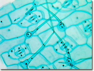
Hanging Drop Method for Bacterial Motility • Microbe Online Hanging Drop Method Preparation. Take a clean glass slide and apply a paraffin ring, adhesive-tape ring to make circular concavity. (This step is not needed if a glass slide with depression is available). Hold a clean coverslip by its edges and carefully dab vaseline on its corners using a toothpick. Place a loopful of the fresh broth culture ... Mitosis in Onion Root Tips - Amrita Vishwa Vidyapeetham Then the thin layer of cell squash on the slide was viewed under the light microscope. Then the cell was photographed and documented. By using actively dividing cells in the onion root tip, this experiment aims to obtain a karyotype from the sample and to determine the purpose of each step used in the procedure. MCQ on Microscopy Pdf - YB Study (1) A microscope is an instrument that can be used to observe small objects, even cells. (2) A microscope is a laboratory instrument used to examine objects that are too small to be seen by the naked eye. (3) Both of these (4) None of these Answer: 3 2. Which microscope is more powerful? (1) Electron microscope (2) Compound Microscope 11 Different Types of Microscopes (With Pictures) - Optics Mag The 11 Types of Microscopes: 1. Light Microscopes. The most common type of microscope you're likely to come across, these microscopes rely on lenses and light to illuminate a specimen for optimal image-gathering. They can be used for viewing living cells, insects, for performing dissections, or for clinical blood and tissue assessment.

Parts Of A Microscope Worksheet | Homeschooldressage.com
5 White Blood Cells Types and Their Functions - New Health Advisor There are two different kinds of white blood cells and each looks different from one another under the microscope. These include granulocytes and agranulocytes. Granulocytes have visible granules or grains inside the cells that have different cell functions. Types of granulocytes include basophils, neutrophils, and eosinophils.

Cell Types and Organelles
Mitosis- definition, purpose, stages, applications with diagram Mitosis definition. Mitosis is the process of cell division in which one cell gives rise to two genetically identical daughter cells, resulting in cell duplication and reproduction. The number of chromosomes is preserved in both the daughter cells. Mitosis is a short period of chromosome condensation, segregation, and cytoplasmic division.
(22).jpg)
An Ultimate Quiz On Microscope Parts And Functions! - ProProfs Quiz
Histology, Astrocytes - StatPearls - NCBI Bookshelf using these tools, astrocytes perform a number of specific functions: neurotransmitter uptake and recycling, promotion of neuronal survival and synapse formation, regeneration of cns damage, propagation of signal transmission between neurons via calcium signaling, potassium ion buffering, maintenance of ph and fluid levels in the brain, removal …
Molecular Expressions: Science, Optics & You - Olympus MIC-D: Brightfield Gallery - Spiderwort ...
Flagella: Structure, Arrangement, Function - Microbe Online Bacterial flagella are long, thin (about 20 nm), whip-like appendages that move the bacteria towards nutrients and other attractants. Flagella are free at one end and attached to the cell at the other end. Flagellum can never be seen directly with the light microscope but only after staining with special flagella stains that increase their ...

31 Drag The Label To The Appropriate Part Of The Microscope. - Labels Design Ideas 2020
Human Biology Lab Online | Lab 2 - Microscopes, Cell Structure and Function The virtual scope has all the same controls found on the real thing. Be sure to go through the complete checklist Microscope controls: turn knobs (click and hold on upper or lower portion of knob) throw switches (click and drag) turn dials (click and drag) move levers (click and drag) changes lenses (click and drag on objective housing)

FISITECH | The Way of Never Give Up: Understanding Standard Laboratory Procedures for Better ...
Sweat glands: Structure and function - Kenhub Eccrine glands are found all over the body and secrete a watery product that cools the skin. Apocrine sweat glands are mainly found in the armpits and perianal area, and secrete a more viscous, odorous product. There are several histological differences between these two types of glands, but they do share a common general structure.

Simple Microscope Labeled Diagram - Micropedia
proscopehr At the end of each round, all bets and winnings are gathered into a central pot. In a nutshell, poker is a game of chance, but it's fun nonetheless. If you're feeling lucky, you'll have a chance to make a nice profit from the game. Poker can be played with any number of players, but the optimal number is six to eight.
Post a Comment for "39 microscope with labels and functions"