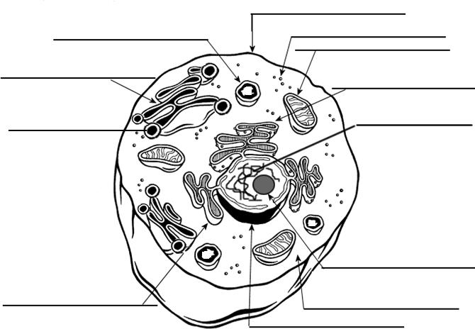
41 diagram of a human cell with labels
YOLO: Real-Time Object Detection Explained YOLO stands for You Only Look Once, and it's an algorithm for real-time object detection and recognition in images. The algorithm was proposed by Redmond et. al in a paper first published at the IEEE/CVF Conference on Computer Vision and Pattern Recognition (CVPR) in 2015. The paper won the OpenCV People's Choice Award. Universal immunotherapeutic strategy for hepatocellular ... Human 293FT cell line used for lentivirus packaging was kept in house and cultured as described previously . All cells except for DCs were grown at 37 °C in 5% CO 2 in DMEM supplemented with 10% exosome-depleted FBS and 1% PS as described above. All the cell lines used were tested to rule out the presence of mycoplasm contamination.
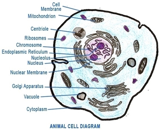
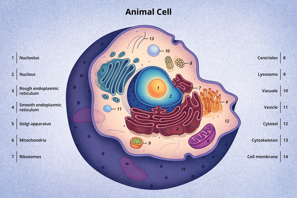
animal cell video free download - costaricavacationtipsh A clear design animal cell diagram template from Edraw is waiting for you in the free download version. An illustration of two cells of a film strip. You can also adjust the diagram sizes at any time you want for more insights. All living organisms are composed of cells from just one unicellular to many trillions multicellular.
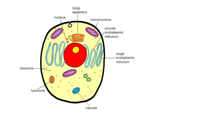
Diagram of a human cell with labels
Cell Membrane (Plasma Membrane) - Genome.gov Definition The cell membrane, also called the plasma membrane, is found in all cells and separates the interior of the cell from the outside environment. The cell membrane consists of a lipid bilayer that is semipermeable. The cell membrane regulates the transport of materials entering and exiting the cell. Cell Membrane 3-D Watch on Narration Skin Cell Labeled - parathyroid glands structure function ... Skin Cell Labeled. Here are a number of highest rated Skin Cell Labeled pictures upon internet. We identified it from obedient source. Its submitted by supervision in the best field. We allow this nice of Skin Cell Labeled graphic could possibly be the most trending subject past we share it in google improvement or facebook. Time‑correlated single‑photon counting technique to ... Time‑correlated single‑photon counting (TCSPC) technique is a powerful way to measure the weak light signals. The basic principle behind TCSPC is the photoelectric effect in which an ...
Diagram of a human cell with labels. STING-Mediated Innate Immunity: How One Discovery Unlocked ... Since it is too early to perform human clinical trials using these newly discovered compounds, Pryde et al. instead exposed C53 to human embryonic kidney cell samples generated in a lab. The ... WHMIS 2015 - Pictograms : OSH Answers Most pictograms have a distinctive red "square set on one of its points" border. Inside this border is a symbol that represents the potential hazard (e.g., fire, health hazard, corrosive, etc.). Together, the symbol and the border are referred to as a pictogram. Pictograms are assigned to specific hazard classes or categories. Cell Cycle - Genome.gov Cell cycle is the name we give the process through which cells replicate and make two new cells. Cell cycle has different stages called G1, S, G2, and M. G1 is the stage where the cell is preparing to divide. To do this, it then moves into the S phase where the cell copies all the DNA. So, S stands for DNA synthesis. Positions and Functions of the Four Brain ... - MD-Health.com The human brain is the most complex organ in the body. Composed of 50 to 100 billion neurons, the human brain remains one of the world's greatest unsolved mysteries. Here we will take a closer look at the four lobes of the brain to discover more about the location and function of each lobe.
Heat Flow and Diagrams Lab - Activity - TeachEngineering Student pairs design, redesign and perform simple experiments to test the differences in thermal conductivity (heat flow) through different media (foil and thin steel). Then students create visual diagrams of their findings that can be understood by anyone with little background on the subject, applying their newly learned art vocabulary and concepts to clearly communicate their results. Learn all muscles with quizzes and labeled diagrams | Kenhub Labeled diagram View the muscles of the upper and lower extremity in the diagrams below. Use the location, shape and surrounding structures to help you memorize each muscle. Once you're feeling confident, it's time to test yourself. Unlabeled diagram See if you can label the muscles yourself on the worksheet available for download below. Male urethra: Anatomy and function - Kenhub The male urethra is an 18-22 cm long muscular tube that conveys urine from the urinary bladder to the exterior via an external opening in the perineum and also functions to provide an exit for semen (sperm) and glandular secretions during ejaculation. It runs from the internal urethral orifice of the bladder to the external urethral orifice located at the tip of the glans penis. Computational profiling of hiPSC-derived heart organoids ... a Diagram of the MULTI-seq experimental workflow. b UMAP projections of the single cells grouped by (i) conditions, (ii) differentiation methods, (iii) cell lines, and (iv) stages. As for all UMAPs...
Circulatory System Diagram - New Health Advisor There are different types of circulatory system diagrams; some have labels while others don't. The color blue stands for deoxygenated blood while red stands for blood which is oxygenated. Below you'll see diagram specified to the heart, as well as circulatory system diagram of the whole body: How Does the Human Circulatory System Work? 1. Heart Anatomical Position Quiz Questions And Answers - ProProfs Here we bring you an anatomical position quiz! The organs in most humans' bodies are located in the same area, and their locations can be described using certain words. As a medical student, you are expected to know what a given term means when it comes to an organ's position. Think you can do this with ease? This quiz is for you to test your understanding. Give it a try! Human Ovary Diagram - testis cartoons illustrations vector ... Human Ovary Diagram - 9 images - file ovary corpus embryology, print exercise 43 pictures flashcards easy notecards, Kidney Structures and Functions Explained (with Picture ... Kidney Structures and Functions Explained (with Picture and Video) Your kidneys are paired organs found on each side of the back portion of the abdominal cavity. The larger left kidney is located a bit higher than the right kidney. Unlike other organs found in the abdomen, the kidneys are located behind the lining (peritoneum) of the abdominal ...
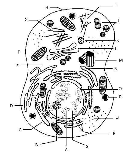
Parts of Cells | Science - Quizizz
Cell Membrane Diagram - solved which of the following ... Cell Membrane Diagram. Here are a number of highest rated Cell Membrane Diagram pictures on internet. We identified it from trustworthy source. Its submitted by management in the best field. We receive this nice of Cell Membrane Diagram graphic could possibly be the most trending topic next we allowance it in google help or facebook.
(SOLVED) HLTAAP001 Recognise healthy body systems
DNA Build - Activity - TeachEngineering DNA: The Human Body Recipe. As a class, students work through an example showing how DNA provides the "recipe" for making human body proteins. They see how the pattern of nucleotide bases (adenine, thymine, guanine, cytosine) forms the double helix ladder shape of DNA, and serves as the code for the steps required to make gene...
Cell | Anatomy System - Human Body Anatomy diagram and chart images
Parts of Human Eye and Their Functions | MD-Health.com Parts of Human Eye and Their Functions Understanding the different parts of our eye can help you understand how you see and what you can do to help keep the eye functioning properly. The eye is one of the most complex parts of the body.

Human digestive system diagram & function explained
Mapping the Brain to Understand the Mind - Scientific American This closeup of a single human neuron highlights just how interconnected brain cells are. False color reveals the locations and abundance of synapses where the cell receives signals from other...

September 2010 ~ Pass. Science. Solutions.
Endothelial Cells Mediated by UCP2 Control the Neurogenic ... Knockdown of endothelial UCP2 accelerates human NPCs differentiation toward astrocytes. A) The diagram of human CMEC/D3 and human NPCs coculture system. B) Schematic of human embryonic stem cells differentiate into human NPCs, followed by neurons and astrocytes. C) RT-PCR analysis showing the knockdown efficiency of human UCP2-shRNA at the mRNA ...

Organisms are composed of cells, and these cells have specific structures within in them that ...
Human neural stem cell-derived extracellular vesicles ... Cell culture. SH-SY5Y cells, a human dopaminergic neuroblastoma cell line, and human foreskin fibroblasts (HFF) were purchased from the American Type Culture Collection (ATCC). F3 cells, a human fetal telencephalon (15 weeks gestation)-derived immortal NSC line, were provided by Prof. Seung U Kim from Chung-Ang university of Korea.

Human cell diagram | school project | Pinterest | Human cell diagram and Homeschool
Hemoglobin - Wikipedia Hemoglobin, (haemoglobin BrE) (from the Greek word αἷμα, haîma 'blood' + Latin globus 'ball, sphere' + -in) (/ ˌ h iː m ə ˈ ɡ l oʊ b ɪ n, ˈ h ɛ m oʊ ˌ-/), abbreviated Hb or Hgb, is the iron-containing oxygen-transport metalloprotein in red blood cells (erythrocytes) of almost all vertebrates (the exception being the fish family Channichthyidae) as well as the tissues of some ...
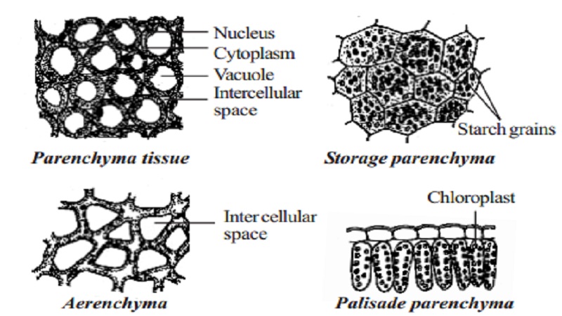
Permanent tissue: characteristics, types and functions - Online Biology Notes
Anatomy of the Epidermis with Pictures - Verywell Health The four layers of cells, beginning at the bottom, are the stratum basale, stratum spinosum, stratum granulosum, and stratum corneum. In your palms and soles, there's an additional layer called stratum lucidum underneath the stratum corneum. In the bottom layer, keratinocytes divide and push up formed cells toward the upper layer.

Labelled Cell | Teaching Resources
Isolation of RNA - Amrita Vishwa Vidyapeetham Growth medium on the cells was discarded and cells were washed with ice cold 1X PBS. The monolayer was then covered with 1 ml of l TRIzol and the cells were lysed and homogenized by repeated pipetting. 2. Phase Separation . The homogenized samples were incubated for 5 minutes at 15 to 30°C for the complete dissociation of nucleoprotein complexes.
Chapter 3 Notes - The Cell
Carcinogenesis - Wikipedia Carcinogenesis, also called oncogenesis or tumorigenesis, is the formation of a cancer, whereby normal cells are transformed into cancer cells.The process is characterized by changes at the cellular, genetic, and epigenetic levels and abnormal cell division.Cell division is a physiological process that occurs in almost all tissues and under a variety of circumstances.
Discovery and Structure of Cells | Biology | Visionlearning
Time‑correlated single‑photon counting technique to ... Time‑correlated single‑photon counting (TCSPC) technique is a powerful way to measure the weak light signals. The basic principle behind TCSPC is the photoelectric effect in which an ...
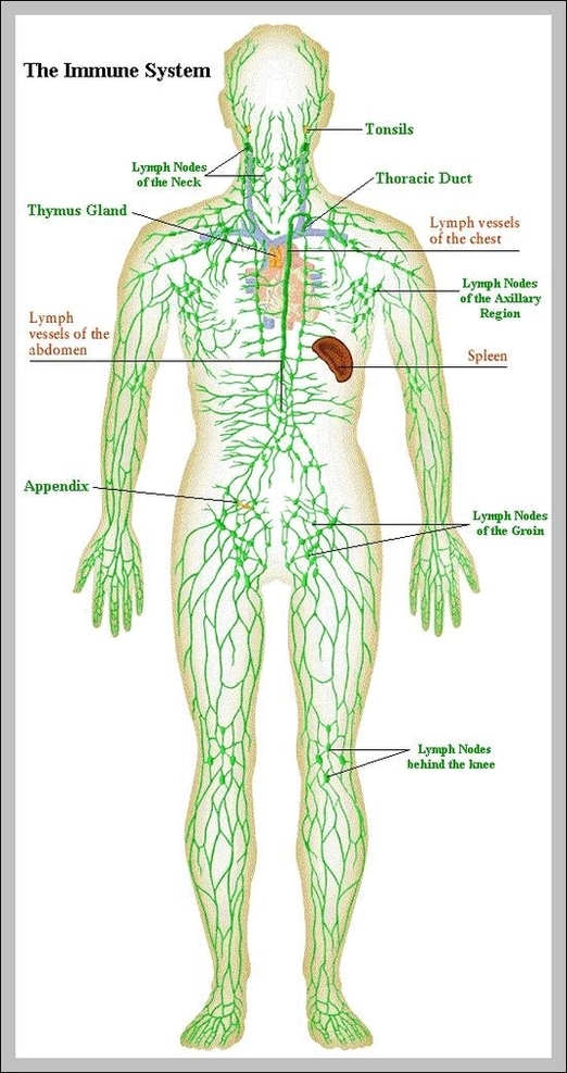
lymphatics | Anatomy System - Human Body Anatomy diagram and chart images
Skin Cell Labeled - parathyroid glands structure function ... Skin Cell Labeled. Here are a number of highest rated Skin Cell Labeled pictures upon internet. We identified it from obedient source. Its submitted by supervision in the best field. We allow this nice of Skin Cell Labeled graphic could possibly be the most trending subject past we share it in google improvement or facebook.

human cell flashcards | Quizlet
Cell Membrane (Plasma Membrane) - Genome.gov Definition The cell membrane, also called the plasma membrane, is found in all cells and separates the interior of the cell from the outside environment. The cell membrane consists of a lipid bilayer that is semipermeable. The cell membrane regulates the transport of materials entering and exiting the cell. Cell Membrane 3-D Watch on Narration

The human egg cell explained for egg donors | Altrui

Mrs. Glaze's 5th grade class: October 2012

female human anatomy organs diagram 2 | Anatomy System - Human Body Anatomy diagram and chart images
Post a Comment for "41 diagram of a human cell with labels"